Copyright © 2022 Foshan MBRT Nanofiberlabs Technology Co., Ltd All rights reserved.Site Map
In recent years, with the rapid development of nanotechnology, nanomaterials have garnered significant attention due to their unique physical and chemical properties. Among these, zinc oxide (ZnO) stands out as an important n-type semiconductor material. It is highly regarded for its direct band gap (3.37 eV), large exciton binding energy (60 meV), and excellent thermal and chemical stability. These characteristics have led to its widespread application in photoelectronic devices, sensors, and photocatalysis. Particularly, ZnO nanofibers, with their high aspect ratio, high electron mobility, and anisotropy, have demonstrated superior performance. However, the properties of ZnO are strongly dependent on the material size. Larger ZnO particles tend to have higher recombination rates of photo-induced carriers, which results in lower photocatalytic activity. Therefore, doping ZnO with transition metal ions such as nickel (Ni) and cobalt (Co) to modulate its physical and chemical properties has become a research hotspot.
Among various methods for fabricating nanofibers, electrospinning with an electrospinning machine has attracted considerable attention due to its simplicity, low cost, and ability to produce continuous nanofibers with uniform diameters. In this study, pure ZnO, Ni-doped, and Co-doped ZnO nanofibers were successfully synthesized via electrospinning, and the effects of doping on the structural, morphological, optical, and magnetic properties of the nanofibers were systematically investigated.
In this experiment, polyvinyl alcohol (PVA) was used as the matrix material. ZnO precursor solutions were electrospun into fibers using an electrospinning device and subsequently calcined at 500°C to remove organic components and form ZnO nanofibers. The doping concentrations of Ni and Co were both set at 5 mol%. The samples were comprehensively characterized using scanning electron microscopy (SEM), X-ray diffraction (XRD), Fourier-transform infrared spectroscopy (FTIR), UV-vis diffuse reflectance spectrophotometer (DRS), and vibrating sample magnetometer (VSM).

Fig. 1. Schematic of the preparation process of samples.
The surface morphologies of pure ZnO and Ni- and Co-doped ZnO precursor nanofibers were observed using SEM. The morphological characterization based on the recorded SEM images indicated that the obtained structures could be classified as nanofiber structures, with fibers exhibiting random orientation and smooth, uniform surfaces. The diameters of the pure ZnO and Ni- and Co-doped ZnO precursor nanofibers were 470 nm, 417 nm, and 426 nm, respectively. After calcination at 500°C, the fiber diameters significantly decreased to 180.7 nm, 133 nm, and 128 nm, respectively. This reduction in diameter was attributed to the removal of PVA from the fiber structure. Additionally, histograms of the fiber diameter distributions further confirmed the changes in fiber diameter (Figure 2).

Fig2 diameter size distribution of fibers, (a) ZnO, (b) Ni-doped ZnO, and (c) Co-doped ZnO.
The crystalline structures of pure ZnO and Ni- and Co-doped ZnO nanofibers were investigated using XRD. The XRD analysis confirmed that the samples possessed a highly crystalline hexagonal wurtzite structure, with no secondary phases detected. As shown in Figure 3, all samples exhibited the hexagonal wurtzite structure (space group P63mc), with multiple characteristic peaks along the (100), (002), (101), (102), (110), (103), (201), and (004) planes. No additional peaks associated with compounds such as Co, Ni, NiO, and Co₃O₄ were observed in the doped samples, indicating that Ni and Co ions were successfully incorporated into the ZnO lattice without forming secondary phases.

Fig 3 The X-ray diffraction patterns of different samples.
Regarding optical properties, the optical absorption behavior of the nanofibers was studied using UV-vis DRS. As shown in Figure 4, the absorption edge of undoped ZnO was located at approximately 370 nm, with a band gap value of 3.18 eV. Ni-doped ZnO nanofibers exhibited a slight blue shift, with a band gap value of 3.20 eV. In contrast, Co-doped ZnO nanofibers showed a red shift, with the absorption edge at 405 nm and a band gap value of 2.82 eV. This variation in band gap was attributed to the size effect and the influence of doping on the lattice structure. Additionally, Co-doped ZnO nanofibers displayed peaks at 568 nm, 610 nm, and 659 nm, corresponding to d–d crystal field transitions in the high-spin state (S = 3/2).

Fig 4 The absorbance spectra of undoped and doped ZnO nanofibers.
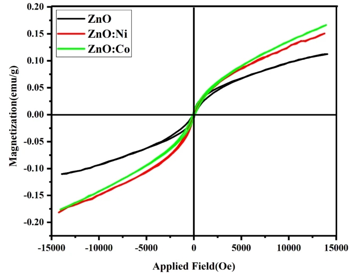
The magnetic properties of the nanofibers were examined using VSM, revealing ferromagnetic characteristics (Figure 9). All samples exhibited ferromagnetic behavior at room temperature. The magnetization of Ni- and Co-doped ZnO nanofibers was higher than that of undoped ZnO nanofibers. This ferromagnetic behavior was attributed to the substitution of non-magnetic elements (Zn) with transition metal ions (Ni, Co) and the presence of intrinsic defects in the lattice.

Fig 5 Magnetization vs. magnetic field curve measured at room temperature of pure and Co, Ni-doped ZnO.
In this study, pure ZnO, Ni-doped, and Co-doped ZnO nanofibers were successfully synthesized via electrospinning machine. The effects of doping on the structural, morphological, optical, and magnetic properties of the nanofibers were systematically investigated. The results indicated that Ni and Co ions were successfully incorporated into the ZnO lattice without forming secondary phases. The morphological characterization based on SEM images confirmed that the obtained structures were nanofiber structures, and the reduction in fiber diameter was due to the removal of PVA from the fiber structure. XRD analysis confirmed the highly crystalline hexagonal wurtzite structure of the samples, with no secondary phases detected. The band gap values of undoped and doped ZnO nanofibers with Ni (5%) and Co (5%) were estimated to be 3.18 eV, 3.20 eV, and 2.82 eV, respectively, influenced by the size effect. The magnetic properties of the nanofibers exhibited ferromagnetic characteristics.
These findings provide important theoretical and experimental support for the application of ZnO nanofibers in photoelectronic devices and sensors. Future work will focus on investigating the effects of different doping concentrations on the properties of ZnO nanofibers and exploring their potential applications in practical devices.
Article Source: https://doi.org/10.1038/s41598-025-95443-7